

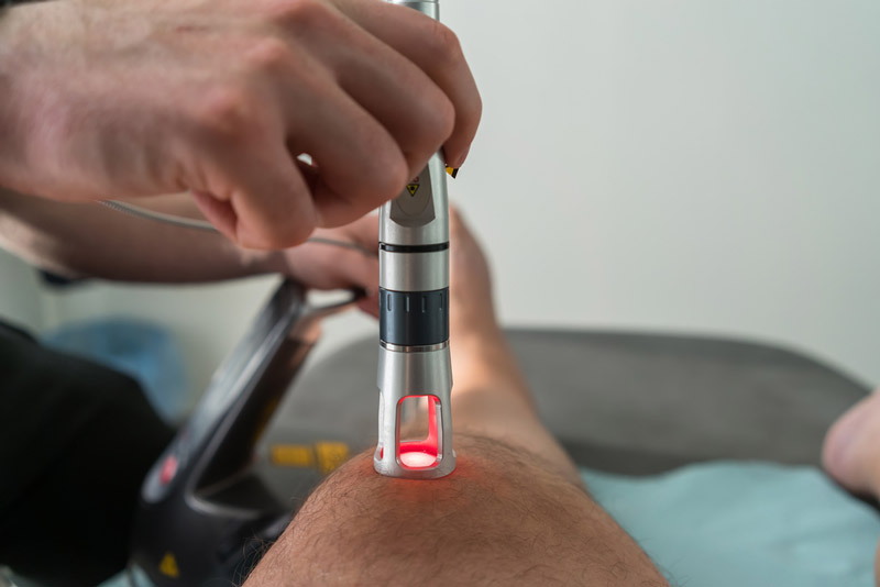
Interventional pain medicine has advanced significantly in recent decades, integrating imaging technologies that enable procedures to be performed with greater safety, accuracy, and patient-specific adaptation. Two of the most widely used modalities for image-guided pain interventions are ultrasound and fluoroscopy. Both are highly effective, but choosing the right one depends on clinical goals, anatomical considerations, and logistical factors. What the procedure involves locally is set out in fluoroscopic guided injections in Brooklyn.
This article explores the technical foundations, benefits, limitations, and clinical applications of each modality, and how they shape high-precision care in settings like advanced pain management centers in Brooklyn.
Technical Overview of Each Modality
Ultrasound-Guided Injections
Ultrasound uses high-frequency sound waves to generate real-time images of anatomical structures. In interventional medicine, its value has grown rapidly due to its ability to visualize soft tissues, blood vessels, nerves, and articular structures, both superficial and moderately deep.
Key Advantages:
-
Real-time, dynamic, 2D imaging.
-
Continuous visualization of the needle in motion.
-
No ionizing radiation exposure.
-
Portable and immediately available at the point of care.
-
Allows pre-, intra-, and post-procedure anatomical assessment.
🔵 Fluoroscopically Guided Injections
Fluoroscopy is a radiological technique that provides real-time imaging through continuous X-ray exposure. It remains the gold standard for many spine procedures due to its excellent visualization of bony anatomy.
Key Advantages:
-
High-resolution imaging of bone structures.
-
Use of contrast dye to confirm precise needle placement or medication spread.
-
Ideal for complex spinal procedures and deep joint access.

In-Depth Clinical Comparison
🎯 Anatomical Precision & Safety
-
Ultrasound enables visualization of key structures like arteries, veins, and nerves, improving safety and reducing the risk of iatrogenic injury, particularly in peripheral interventions.
-
Fluoroscopy, while unable to display soft tissues directly, excels when bone alignment is critical, as in spinal or sacroiliac joint procedures.
👁️ Dynamic Imaging Capabilities
-
With ultrasound, clinicians can adjust the needle trajectory in real time in response to tissue resistance, motion, or drug dispersion.
-
Fluoroscopy offers live imaging but is limited in its ability to visualize three-dimensional relationships outside of bone and contrast-filled spaces.
☢️ Radiation Safety
-
A major advantage of ultrasound is zero radiation exposure, which is critical for vulnerable populations (e.g., children, pregnant patients) or those requiring repeated interventions.
-
Fluoroscopy involves ionizing radiation, typically low-dose but cumulative over time, impacting both patients and practitioners.
⏱️ Efficiency & Workflow
-
Ultrasound supports streamlined workflows: faster setup, no radiation shielding needed, and use in outpatient clinics or bedside environments.
-
Fluoroscopy requires specialized equipment, protective measures, and typically a controlled setting such as a procedure suite or OR.
Clinical Application by Region
🦴 Spinal Procedures (Best with Fluoroscopy)
-
Epidural steroid injections (transforaminal, interlaminar, caudal).
-
Facet joint and medial branch blocks (cervical, thoracic, lumbar).
-
Sacroiliac joint injections, especially in patients with complex bony anatomy.
Fluoroscopy offers unmatched accuracy when precise needle alignment with vertebral landmarks is essential.
🦵 Peripheral Joints and Superficial Nerves (Best with Ultrasound)
-
Joint injections (shoulder, hip, knee, ankle, wrist).
-
Peripheral nerve blocks (femoral, sciatic, radial, etc.).
-
Soft tissue conditions (bursitis, tendinopathies, plantar fasciitis).
Ultrasound allows direct visualization of inflammation, fluid, fibrosis, and tissue pathology, features that X-ray alone cannot detect.
Evidence & Evolving Trends
Numerous clinical studies and systematic reviews have compared ultrasound and fluoroscopic guidance in terms of precision, pain relief, and functional outcomes. Key findings include:
-
Ultrasound-guided joint injections often achieve clinical success equal to or greater than fluoroscopic approaches, especially in peripheral joints.
-
For spinal interventions, fluoroscopy remains the most validated modality, although ultrasound is gaining ground in certain paravertebral and cervical blocks.
Moreover, growing concerns over radiation exposure are driving wider adoption of ultrasound in settings where patients require ongoing pain interventions.
In Practice: Technology in the Service of the Patient
Pain management centers that offer both technologies can match the intervention to the specific condition rather than to the equipment on hand. The guiding principle is simple: not every tool is right for every patient, but every patient deserves access to the right technology.
Options include:
-
Ultrasound-guided injections: radiation-free, real-time soft tissue precision.
-
Fluoroscopically guided spinal and facet injections: accurate to the millimeter with radiological confirmation.
🎯 Conclusion
Choosing between ultrasound and fluoroscopic guidance should never be based solely on convenience or habit. It should be a deliberate, evidence-based decision, tailored to the patient’s anatomy, the clinical indication, and the goals of care.
In precision pain medicine, accuracy is not optional, it’s essential.
A dual-modality imaging strategy supports better clinical outcomes, enhanced safety, and a better patient experience.
Frequently asked questions
Can a patient ask for ultrasound instead of fluoroscopy to avoid radiation?
It is a reasonable question to raise, and for many peripheral joint and soft tissue injections ultrasound is already the preferred choice. For most spinal procedures fluoroscopy remains the validated method, because needle position against bone and the spread of contrast cannot be confirmed any other way. Where both are technically suitable, patient preference is a fair part of the discussion.
What is the contrast dye used in a fluoroscopic injection?
It is an iodine-based agent injected in a small amount so the spread of medication can be checked before the drug itself is given. Reactions are uncommon but possible, and any previous reaction to contrast media should be mentioned in advance. Alternatives include a different agent or, in some cases, completing the procedure without contrast. Kidney function is also considered where relevant.
Does using image guidance make an injection take longer or hurt more?
Not usually. Ultrasound adds a scanning step but no extra needle passes, and often reduces them, since the needle can be steered on the first attempt. Fluoroscopy involves positioning and a contrast check, so the appointment runs longer, though the injection itself is comparable. Most of the discomfort comes from the needle and the volume injected rather than from the imaging.
Does body size affect which type of imaging guidance is chosen?
It can. Ultrasound loses detail with depth, so a deep target in a larger patient may be hard to see clearly, and fluoroscopy or CT can give a more reliable view. The reverse also applies, since superficial nerves and small joints are usually easier to see with ultrasound. The decision is made on the specific target and on the quality of the images obtained.
Unsure whether an injection is the right next step?
A physician-led evaluation weighs it against the alternatives. More on ultrasound guided injections.
Serving Brooklyn, the Five Towns, and Monsey.


