Ultrasound Guided Injection
Ultrasound-Guided Joint, Hip, Shoulder Injections In Brooklyn, NYC
What Is An Ultrasound-Guided Injection?
Ultrasound-Guided Injections are the latest advancement in pan management. This technique administers medications to the painful area using ultrasound guidance. Ultrasound is a device that provides live imagining of the structures in your body and the needle’s position as it penetrates your tissues.
Ultrasound guidance prevents the need for a hit-or-miss approach because it guides the needle directly at the source of your pain. It’s an advanced injection therapy that is minimally invasive, safe and allows extreme accuracy when delivering the medication.
We administer cortisone, lidocaine, or hyaluronic acid with ultrasound to provide pain relief and tissue healing. The ultrasound-guided injection treats various musculoskeletal pain by reducing inflammation and promoting your natural healing response:
- Cortisone injections: Ultrasound-guided cortisone injections reduce inflammation and prevent further damage from acute and chronic musculoskeletal injuries.
- Lidocaine injections: Lidocaine numbs pain receptors and blocks the transmission of pain signals to your brain and spinal cord. It’s a local anesthetic that will leave you pain-free for days or weeks.
- Hyaluronic acid (HA) injections: Also known as hyaluronan, HA lubricates your joints to reduce painful friction. It also functions as a shock absorber for heavy loads acting on your joints. It’s an excellent alternative to knee replacement surgery due to osteoarthritis.
Leading pain management specialist Dr. Henry Sardar expertly performs ultrasound-guided injections right here in Brooklyn, New York. Visit PainTherapy Medical Care to book your session and benefit from our state-of-the-art facilities.
How Do Ultrasound-Guided Injections Relieve Pain?
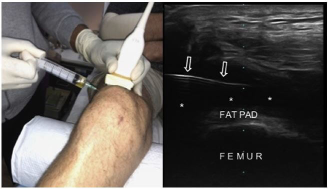
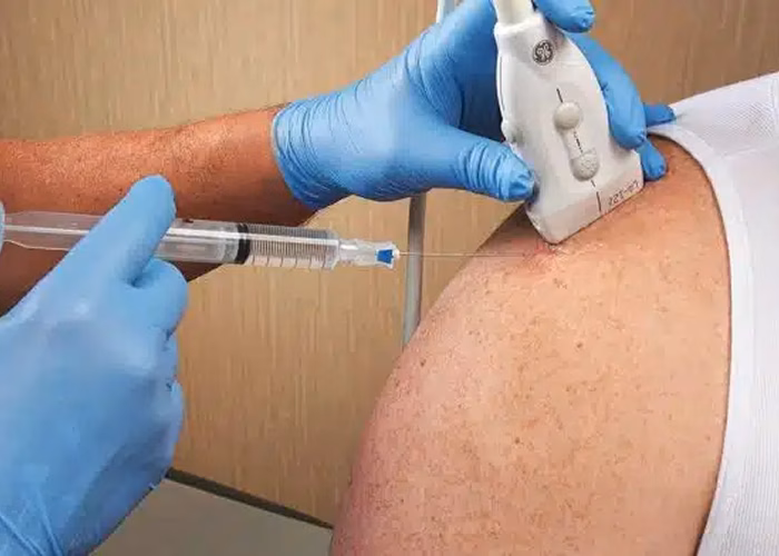
Using the ultrasound, Dr. Sardar performs a preliminary scan of the affected area to locate the precise location to insert the needle. Once the site is located, he preps your skin with an antiseptic solution.
Typically, ultrasound-guided injections use a mixture of steroids and local anesthetic to numb the pain and soothe the area. We hold the ultrasound probe over the target area, and Dr. Sardar pushes the needle into your skin and toward the painful tissue. The ultrasound’s real-time imaging allows him to avoid nearby structures and administer the injection with precision.
It’s an outpatient procedure that requires very little preparation or post-appointment support. The entire process can take up to 5 to 10 minutes. After a short monitoring period, you’re free to go right after the procedure. The anesthetic effect of the ultrasound-guided injection can last days after your session. Evidence from clinical trials reveals significant long-term improvements up to 6 weeks after the injection in some patients.
Can Ultrasound-Guided Injections be Combined with Other Procedures?
At PainTherapy Medical Care, you can combine ultrasound-guided injections with other pain management treatments. Dr. Sardar can devise a tailored treatment plan for your specific condition.
Some therapies that Dr. Sardar often combines with ultrasound injecctions include:
- Cold laser therapy
- Shock-wave therapy
- Cryotherapy
- Kinesiology taping
- TECAR Therapy
- Aqua therapy
- Perineural Injection Therapy (PIT)
- Physical and Occupational Therapy
- Electromagnetic Transduction therapy (EMTT)
- Fluoroscopically Guided Epidural Steroid Injection and Facet Injection
Ultrasound-guided injections bring about localized therapeutic effects to promote your natural healing mechanism. Their ability to trigger tissue regeneration ensures long-term healing and faster recovery. You can now access Ultrasound-Guided Injections at PainTherapy Medical Care. Visit us today to get long-lasting relief from your chronic pain!

What Conditions Do Ultrasound-Guided Injections Treat?
Ultrasound-guided injections are a highly effective therapy for musculoskeletal pain, inflammatory injuries, nerve impingement, and various painful conditions that can affect your overall quality of life. Here are common conditions that Dr. Sardar often treats with ultrasound-guided injection:
- Osteoarthritis
- Ankle joint pain
- Epicondylitis
- Tendonitis
- Tennis elbow
- Morton’s neuroma
- Carpal tunnel syndrome
- Frozen shoulder
- Subacromial bursitis
- Trochanteric bursitis
- Knee Osteoarthritis
- Arthritic joints of the hand and fingers
Feel free to check with our Pain Management doctors what other musculoskeletal conditionsare included in this list.
If you have any of the conditions mentioned above, then it’s time to try out ultrasound-guided injections for fast and effective pain relief. Book an appointment with our pain management doctor at PainTherapy Medical Care in Brooklyn, NYC!
Where Can I Get ultrasound-guided Injections?
Dr. Henry Sardar offers Ultrasound-Guided Injections right here in Brooklyn. Dr. Sardar’s patients in New York can attest to the efficacy of ultrasound-guided injections as a safe treatment that relieves chronic pain and improves the quality of life. Ultrasound guided injection therapy is likely to be covered by insurance if your primary pain management doctor recommends it.
We accept most major insurance providers at our Brooklyn pain clinic: New York State Medicaid, HealthFirst, Fidelis Care, Affinity Group, Metro Plus, Amerigroup, BlueCross / BlueShield Medicaid Plan, and UnitedHealthCare Community Plan. Reach out to us and book an appointment at Pain Therapy Medical Care, New York’s top pain management therapy clinic.